 Rate List MRI Scanning
Customer Support
9109988973, 9109988974
Opening hours
Customer Support
Opening hours

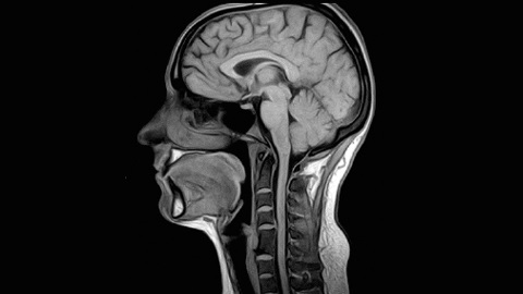
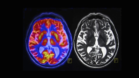
A Brain MRI can help determine whether you sustained any damage from a stroke or head injury. Therefore, the doctor suggests for head MRI. The head MRI is a painless, non-invasive test that generates detailed images of the brain and brain stem. Our Signa HD 3.OT AND Signa HDX 3.OTMRI machine creates the images using a magnetic field and radio waves. The patient with stroke and head injury visits us for their head MRI.
Our Signa Hd and HDx machine provide digital, high-definition, anatomically optimized imaging.
MRI of the brain can be useful in evaluating problems such as persistent headaches, dizziness, weakness, and blurry vision or seizures, and it can help to detect certain chronic diseases of the nervous system, such as multiple sclerosis.
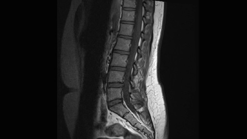
Spine MRI is done using radio waves, a magnetic field and a computer. It creates clear, detailed pictures of the spine and surrounding tissues. MRI does not use radiation and may require an injection of gadolinium contrast material.
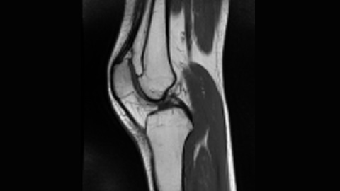
MRI of a knee is a strong magnetic field, which uses radio waves and a processor to generate detailed pictures of the structures within the knee joint. Knee MRI does not use ionizing radiation, and it can help decide whether you require surgery.
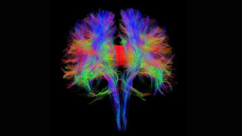
In neuroscience, tractography is a 3D modelling technique used to visually denote nerve tracts using data collected by diffusion MRI. It uses special techniques of magnetic resonance imaging (MRI) and computer-based diffusion MRI. The results are displayed in two - and three-dimensional images called tractograms.
Functional magnetic resonance imaging (fMRI) measures the minute changes in blood flow that occur with brain activity. It may be used to examine the brain's functional anatomy, evaluate the effects of a stroke or other disease, or to guide brain treatment.
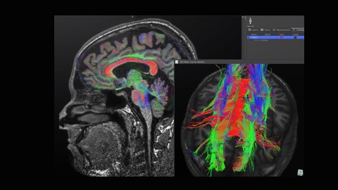
Diffusion tensor imaging (DTI) is a promising technique for identifying microstructural changes or differences with neuropathology and treatment. The diffusion tensor may be used to characterize the magnitude, the degree of anisotropy, and the orientation of directional diffusion.
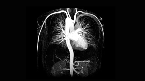
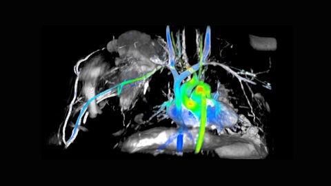
It is a type of MRI that looks particularly at the body's blood vessels.It requires inserting a catheter into the body, magnetic resonance angiography is a far less invasive and less painful test.
While magnetic resonance angiography,you lie flat inside the magnetic resonance imaging scanner. This is a large, tunnel-like tube. In some cases, a special dye, known as contrast, may be added to your bloodstream to make your blood vessels easier to understand.
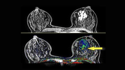
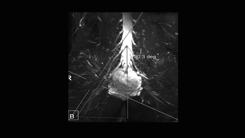
It is fundamentally used as a supplemental means for breast screening with mammography or ultrasound. It may be used to screen women at high risk for breast cancer, assess the extent of cancer following diagnosis, or further assess anomalies seen on mammography. Breast MRI does not use ionizing radiation, and it is the most reliable method for determining whether silicone breast implants have ruptured.
MR Imaging of Peripheral Nerves (PNI), is an advanced technique that helps diagnose disorders of the peripheral nerves beyond the spinal canal. We at Life Imaging Centre provide diagnostic magnetic resonance imaging of the spine, brain, pelvis and extremities.
MR neurography can image nerves everywhere in the body, although it is most commonly used in the diagnosis of abnormalities of the brachial plexus, lumbosacral plexus, thoracic outlet, and sciatic nerves.
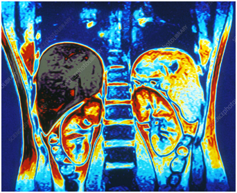
Cardiac MRI is done to provide detailed information on the type and austerity of heart disease which helps the doctor to decide the best approach to treat heart problems such as coronary heart disease, heart valve problems, pericarditis, cardiac tumours, or damage from a heart attack. Cardiac MRI can help in interpreting outcomes from other imaging tests such as chest x - rays and chest CT scans.
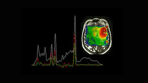
MR spectroscopy is a noninvasive symptomatic test for measuring biochemical differences in the brain, especially in the presence of tumours. While magnetic resonance imaging MRI. It recognises the anatomical location of a tumour, MR spectroscopy compares the chemical composition of normal brain tissue with abnormal tumour tissue.
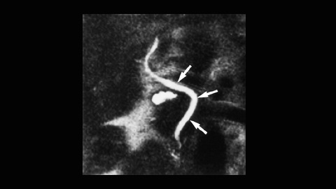
Magnetic Resonance Cholangiography is a fast, accurate, non-invasive option to endoscopic retrograde cholangiography (ERC) in the evaluation of biliary tract disease. Technical advances in imaging sequences and the use of phased-array coils allow high-quality imaging comparable to that available with ERC. In choledocholithiasis, popular bile duct stones as small as 2 mm can be detected with MR cholangiography and look as low-signal-intensity foci within the high-signal-intensity bile. MR cholangiography may help establish the diagnosis of cancerous obstruction and is useful in the evaluation of patients in whom ERC was destroyed or incomplete.
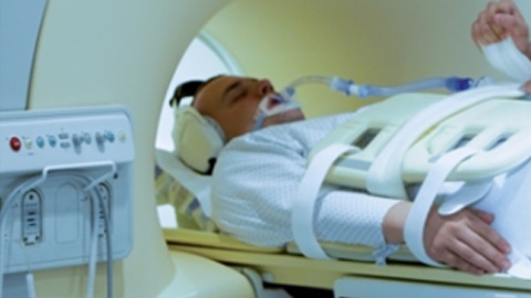
Our fully - equipped ICU ventilator guarantees uncompromised continuous ventilation care from the ICU to the MRI scanner and back. Its reliability and high performance, with advanced lung-protective strategies and patient-adaptive modes, makes us the ideal choice for any critical care department that needs to transport ventilated patients to the MRI department.
The integrated high-performance turbine enables the machine to be completely independent of compressed air. This lessens weight and saves travel time during transport.
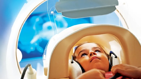
Pediatric magnetic resonance imaging uses a powerful magnetic field, radio waves and a computer to produce accurate pictures of the child's body. Pediatric MRI is used to diagnose and monitor treatment for a variety of conditions within the brain, chest, abdomen, pelvis and extremities.
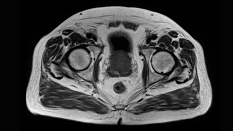
Prostate MRI is used to evaluate the volume of prostate cancer and also concludes whether it is spreading. The doctors use this MRI to diagnose infection and the conditions you were born with, it also detects an enlarged prostate. Some exams may use an anorectal coil, a thin wire covered with a latex balloon. The operator inserts the coil from a short distance into the rectum. Prostate MRI does not use radiation. It provides images that are more precise and more detailed than other imaging methods.
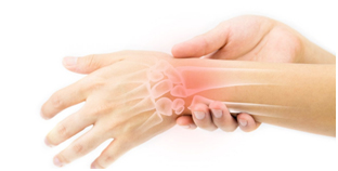
When do we need an MRI scan of the wrist:
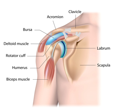
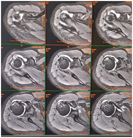
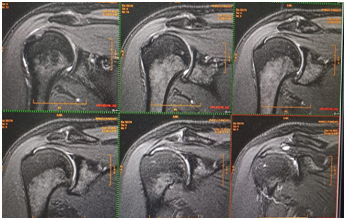
A shoulder MRI helps your doctor diagnose potential problems found in other imaging tests, such as X-rays. It also helps your doctor diagnose unexplained pain in the area or better understand the condition causing your shoulder symptoms. An MRI scan uses no radiation and is considered a safer alternative, especially for pregnant women and children.
Abdominal MRI scans are used for a variety of reasons. Your doctor will order an MRI if they suspect something is wrong in your abdominal area but can’t determine what through a physical examination.
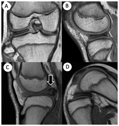
An MRI lets your doctor see the soft tissues in your body along with the bones. This allows them to inspect the elements of the knee that might have been injured during physical activity or from wear and tear. The test can also provide detailed images of various sections of the knee, such as bones, cartilage, tendons, muscles, blood vessels, and ligaments.
MRI is ideal for diagnosing Sport Injuries or other injuries to the Knee
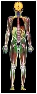
A Whole-Body MRI or also called a Full Body MRI is multiple scans of the entire body with an aim to diagnose any malfunctions inside the body.
It allows for to detection of any early-stage disease or growing pathological condition, and the treatment to that is possible.
The whole-body MRI scan is useful for a routine check-up of an individual who does not have any symptoms.
A whole-body scan aims to identify any potential inflammation, tumour, obstructions, etc.
The whole-body MRI takes about an hour to complete the scan with multiple intervals to ensure that the setup of the equipment is necessary
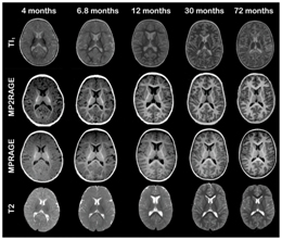
Our specialised MRI facilities are designed for children in order to get thehighest quality images in the shortest amount of time. Our MRI technologists are fully experienced in working with children of all ages with a wide range of conditions.